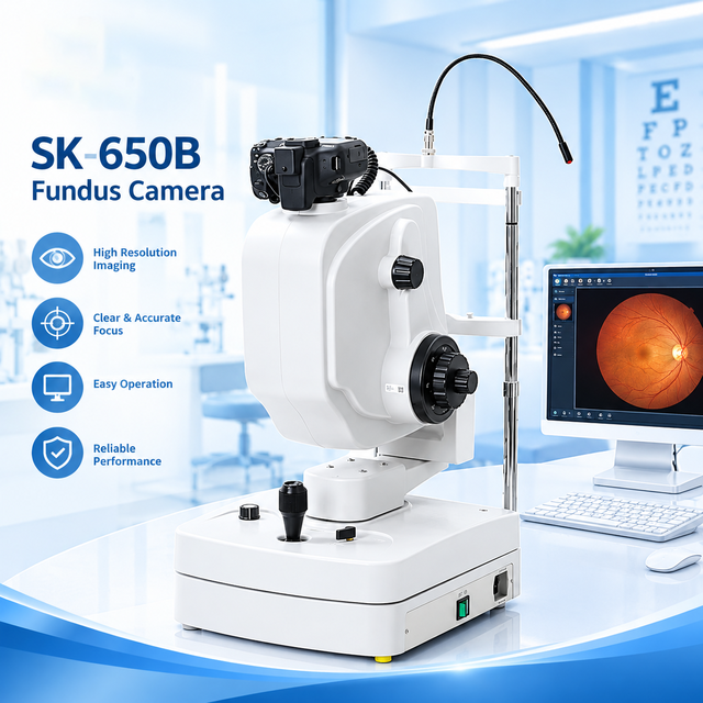
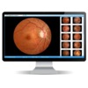
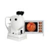
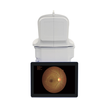
The SK-650B is a non-mydriatic fundus camera designed for digital retinal observation and imaging. Its optical system provides a 45° maximum field of view, while the digital imaging system supports high-resolution color fundus photography and monochrome imaging.
Full-circle optical design provides a maximum 45° field of view for fundus observation.
Designed for non-mydriatic fundus observation and digital retinal imaging.
Supports 5184 Ã 3456 color image resolution for detailed digital fundus photography.
Internal fixation targets assist with patient positioning during fundus examination.
The SK-650B incorporates an aspheric optical system and a movable observation platform. The optical design is intended to provide clear fundus observation while maintaining a practical working distance for clinical examination.
Optical Lens: Aspheric System
Maximum Field of View: 45°
Working Distance: 40 mm ± 2 mm
Compensation Range: -25D to +30D
The digital imaging configuration supports detailed color fundus photography and monochrome imaging for professional ophthalmic observation and documentation.
Resolution: 5184 Ã 3456
Digital camera system based on Canon EOS technology.
Supports digital image processing and patient profile management.
The split-line aligning focus method helps indicate focus status during fundus image acquisition and supports accurate focusing.
The system can identify retinal status and adjust exposure intensity according to the examination condition.
Fundus observation position indication assists with image positioning during examination.
The software supports digital image processing, patient profile management and integration with clinical information systems.
⢠Ophthalmology clinics
⢠Optometry and eye care centers
⢠Hospital ophthalmology departments
⢠Retinal imaging and fundus observation
⢠Digital ophthalmic image documentation
⢠Clinical image management systems
| Model | SK-650B |
| Optical Lens | Aspheric System |
| Test Mode | Non-Mydriatic Fundus |
| Minimum Pupil Size | 3.3 mm |
| Focus Mode | Manual, Split-Line Aligning Focus |
| Capture | Manual |
| Color Image Resolution | 5184 Ã 3456 |
| Compensation Range | -25D to +30D |
| Maximum Field of View | 45° |
| Fixation Target | 9 Internal Fixation Targets |
| Optical Head Tilting | Horizontal ±30°, Vertical ±12.5° |
| Working Distance | 40 mm ± 2 mm |
Digital Fundus Imaging for Professional Ophthalmic Applications
Processing time
According your order list. we will supply delivery time .
Commonly is 5-7 work days after we confirm payment
Shipping time
By Express : 3-7 days . Door to door service
By Air : 3-7 days . (the goods arrival at your local air ports
By sea : 30-40 days (the goods arrival at your local sea ports )
PS : by air or by sea, if you need door to door service, please tell us . we will help you checking the extra door to door cost.
How many months warranty do you provide ?
one year
What should I do if the machine have question ?
For ensuring all the items are in good quality, we will test all products before the packages were sent out. Once the machine have some question, please don't worry. kindly send us some images of the situation , Video and machine series, and explain the question in where . we will supply solution to you deal soon.
What should I do if the spare parts is broken in Warranty ?
If in Warranty, some spare parts broken in the Use. kindly supply picture or video , and machine series number .
We will send the spare parts by express to you soon.. and we bear express cost .
Please enter your target price
Please provide a quantity
United States
Please provide your name
Please enter your email adress
Enter a valid email address
Please enter the phone number
Please enter the inquiry information
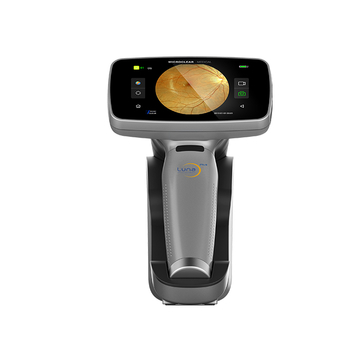
12 million pixel Hand-held fundus camera for ophthalmology
High pixel Hand-held fundus camera
Ophthalmic Instrument Retinal Fundus Camera RET-3100B
LINK-PP LPJ1075-1BHNL 100% Cross RTA-110AHQ1A UDE
United States
Carrier
CostDelivery TimeTracking
SK-650B Non-Mydriatic Fundus Camera 45° Digital Retinal Camera High Resolution Ophthalmic Fundus Imaging System with DICOM