manufacture refraction instruments, ophthalmic diagnostic instruments, visual acuity chart, ophthalmic unit, electric table,one stop medical instrument
-
Optometry Equipments (758)
- Combined Table and chair (120)
- Motorized/Electric Table/Motorized Lift Table (34)
- Electric Chair (33)
- Auto Refractometer/Keratometer (185)
- Handheld /Portable Auto Refractometer (6)
- Auto Phoropter (36)
- Manual Phoropter (27)
- Auto Lensmeter (39)
- Manual Lensmeter (17)
- LCD Chart Vision Monitor (24)
- Auto Chart Projector (12)
-
Optical Lab Machinery (585)
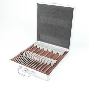
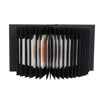
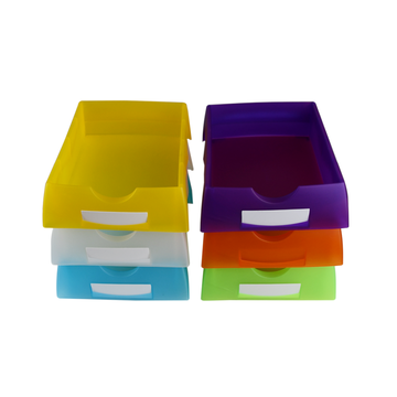
- Trial Lens Frame (80)
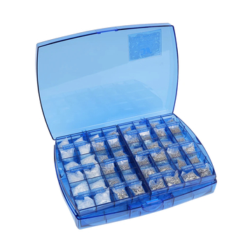
- Trial Lens Set (70)
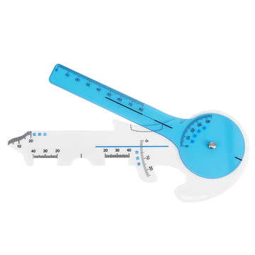
- Pupil Meter/Ruler/PD Meter (65)
- Auto Lens Edger (77)
- Hand Lens Edger (42)
- Lens Cutting Machine (4)
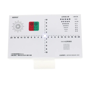
- Visual Acuity Chart (22)
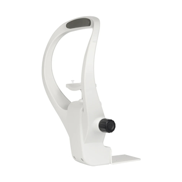
- Phoropter Arm (5)
- Pattern Drilling/Lens Drilling Machine (29)
- Pattern Maker/Optical Lens Pattern Maker (10)
- Lens Groove/Grooveing Machine (22)
- Lens Polish/Auto Polisher (12)
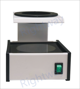
- Frame Heater/Frame Warmer (41)
- HFC (4)
- Ultra Glass Cleaner (30)
- Screw Extractor (6)
- Center Machine (8)
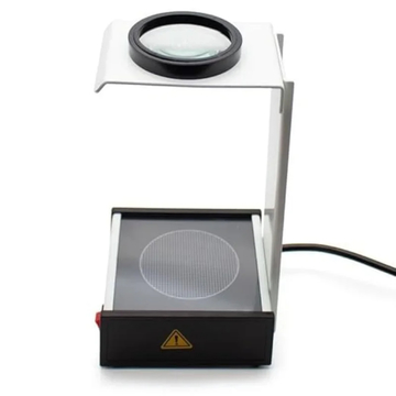
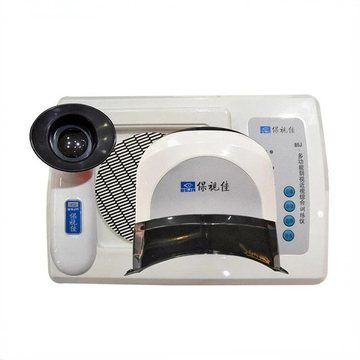
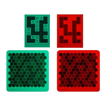
- UV Tester (26)
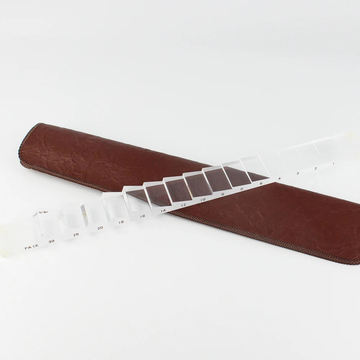
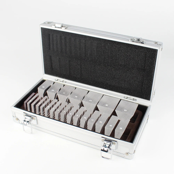
- Lenses Optical Retinoscopy Rack Lens set (19)
- Eyeglass Lens Drilling & Slotting Grooving Machine (4)
- Eyeglasses Screw Extractor (3)
- Layout Blocker (1)
- Lens Tester (1)
- Eyeglass Tools and Spare parts (359)
- Medical Equipment (182)
- Ophthalmic Equipment (47)